The previous article mentioned a lot of cases of fixing fractures in the bone panel. This article will refer to another veterinary fracture internal fixation method-intramedullary needle with ring tie steel wire fixation.We use this technology to fix the long diagonal fracture of the middle end of the cat.
The fixing of the bone panel and the screw is a very popular method in the veterinarian. It can be applied to any fractures of any long bone, especially the middle shaft. It has less pain after surgery and the functional recovery is fast. The screw can be compressed at the fracture, which can increase the friction between the bone breaks and resist the weight of the fracture. It can resist the bending force in the humerus, femoral and tibial fractures, but compared to the bone plate, its anti -axis pressure and anti -rotation ability are poor. The intramedullary needle can only use the friction between the needle and the bones to resist the resistance. Rotating load and axial pressure, usually this friction does not stop the rotation at the fracture and the broken shaft break, so its use still has certain limitations, because there are many problems with many fractures. Combine with ring tie wires to fix the fracture. Now there are some surgeons using two or more intramedullary needles to fix and achieve better results. In the case of this chapter, according to the actual needs, we still chose the intramedullary needle with the ring tie -tie steel wire as the best internal fixed solution.
American short -haired cat American Short Hair
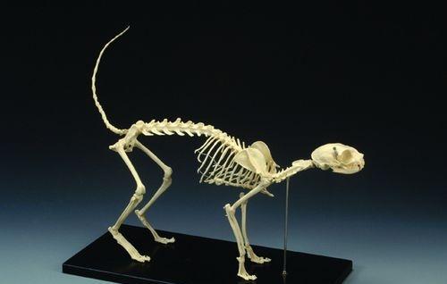
The following cases, the name of the cat with cats: small M, variety: American short -haired cat, age: March age, he was sent to the hospital for treatment from upstairs because of playing, no internal bleeding, etc.EssenceAfter shooting X -rays, there were three fractures of the whole body, which were the left humerus fractures, the neutral fractures of the right rear limb femur and the left hip acetabular fracture.The picture below shows the cat's bone anatomy, and the red arrow shows the position of the humerus.

The picture shows a schematic diagram of cat bones.
Below is Preoperative X -ray Map
The arrow indicates the broken humerus, which is a typical oblique fracture. The surrounding tissue is swollen due to the dislocation of the broken bone.
The arrow indicates the near -end oblique fracture of the femur, and the acetabular fracture can be seen on the opposite side of the arrow
Because the cat is relatively young, the bone growth is fast and the recovery ability is strong, it can heal itself through the fracture of the acetabular nest of the acetabular acetabular nest. Because the three fracture surgery at the same time is very damage to the body, it is determined that only the femoral and the femoral bones and the femoral bones andThe humerus is performed and conservative treatment for the acetabular nest.
Surgical process : Anti -inflammatory, hematopoietic needle, Atto, induced anesthesia after 15 points, and then use inhalation anesthesia.The cat is stable on the side, and the affected limbs are up.The suspension of the affected limb is convenient for surgical operations.Disinfection and isolation of the Department of Surgery.From the rear edge of the humerus nodules to the remote edge of the outer edge to make a skin incision.Do everything along the forward curve of the humerus.Cut the subcutaneous fat and deep fascia, carefully separate and protect the head veins.If necessary, ligate the head vein.Cut the ligament of the arm along the edge of the head muscle and the outer side of the bougainvillea head, be careful of the radial nerve. Once the nerves are separated, the head muscles are exposed, the pectoral muscles and the arm muscles are exposed, and the cut of the humerus is revealed.
Because the cat individual is small, there is no suitable bone plate, and it belongs to a typical long oblique fracture, so the technique of internal intramedullary needle with the steel wire rings is used for fixation.First, the humerus reset, the semi -tie steel wire and the full -ring steel wire are buried in advance.In the center of the bones, the wire is tightened after fixing.When close the incision, suture the surface muscle of the arm head muscle and the arm muscle ligament.Routine suture subcutaneous tissue and muscle.
The picture shows the postoperative X -ray diagram. The humerus is basically returned to normal form, but the fracture line is not aligned. In the later stage, it can be slowly healed by forming a skeleton.
Because the femoral point is almost broken to the large rotary, if the upper part of the intramuscular needle is adopted, there is no enough point of focus, and the fixed effect is not satisfactory.
L -shaped bone connection board used during surgery
X -ray chart fixed with bone screws and bone panels
The postoperative anti-inflammatory was 5-7 days, and the wire was disassembled for two weeks. After the operation, it was brave and restricted to exercise for more than three weeks after surgery.
