The previous article has introduced many fractures of the diaphysis, such as the fracture of the tibial and femoral diaphysis. The surgical method used is the internal fixation of the bone plate. This case belongs to the fracture of the epiphysis, which is close to the growth plate.
In juvenile animals, the fracture of the growth part accounts for about 30% of the fractures, because the bone of the growth part is weaker than the surrounding bones and ligaments.
skeleton diagram
The picture is a picture from a foreign language, which describes the structure of the anterior segment of the tibia in the juvenile (left) and adult (right), the large bone is the tibia, the small is the fibula, and the structures of the tibia from top to bottom are: epiphysis, growth
Fractures of the outgrowth of long bones may occur in the cartilaginous growth plate or tibial tuberosity proximal or distal to the tibia.

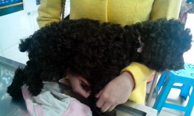
injured dog
The picture shows the dog is limping on the right hind limb and unable to touch the ground
Name of the affected dog: Xiao Q, Age: 5 months old, Breed: Brown Teddy The owner brought him to the hospital for examination because his hind legs could not stand normally after falling from a height.
Affected dog small Q
The patient dog, Xiao Q, is nervous due to pain, and can only be approached when the owner is present
x-ray
Fracture anteroposterior radiograph, the arrow indicates that both the proximal tibia and the fibula were fractured
Palpation of the proximal tibia fracture, the X-ray diagnosis results are shown in the figure below, and the frontal radiograph shows the fracture of the tibial growth part. According to experience, external fixation is more suitable and less damage to the dog and will not affect the function of the growth part, but the lateral radiograph
x-ray
Lateral X-ray shows dislocation of the epiphysis, which can be compared with the image below
x-ray
The arrow shows the lateral view, and there is obvious anterior and posterior dislocation of the fracture part.
Operation preparation: Biochemical examination and blood test were performed before operation. All indicators were normal except for mild anemia. An indwelling needle was placed for preoperative infusion, anti-inflammatory drugs, atropine, and hemostasis were injected.
Surgical procedure: The affected dog was restrained in supine position, the affected limb was elevated, and the surgical skin was prepared, isolated and disinfected.
x-ray
As indicated by the arrow, the crisscross Kirschner wire has basically fixed the proximal tibia
Postoperative care: Routine anti-inflammatory, analgesia, removal of skin sutures one week, and X-ray examination at 4-6 weeks to evaluate the degree of fracture healing.
Because the operation was just done, and the pet owner has not brought it for re-examination, there is no way to know the recovery after the operation. I hope the dog can walk as soon as possible.
