
Diaphragmatic hernia is an abnormal symptom in which the abdominal organs protrude into the thoracic cavity through the diaphragm.
Figure 1 Affected dog
I. Basic situation of the case
The dog was an 8-month-old female Cocker Spaniel, weighing 4 kg, with good nutritional status and good health before the onset of symptoms.
II. Clinical examinations
Body temperature: 37.8 degrees Celsius, heart rate: 185 beats/min, respiration: 75 beats/min, pale oral mucosa, no heart sound on left chest auscultation, weak pulse, and difficulty in breathing.
III. Routine blood biochemical examinations
Table 1 Routine blood test
| items | Results | Reference value | Units |
| RBC | 4.50 | 5.5~8.0 | ×1012/L |
| HCT | 0.25 | 0.37~0.53 | L/L |
| HGB | 83 | 120~170 | g/L |
| items | Results | Reference value |
| Glucose GLU | 15 | 4.5~8.5 |
| Alanine aminotransferase | 188 | 8~75 |
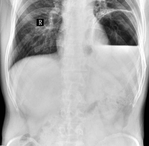
Figure 2 X-ray inspection map
Figure 2 shows that the density of the middle and posterior parts of the right thoracic cavity increases, the cardiophrenic angle disappears, the outline of the diaphragm disappears, and the position of the air-filled bowel in the abdominal cavity moves forward.
According to the results of the above examinations, it was confirmed that the dog had a diaphragmatic hernia due to a car accident.
Considering that the dog's current physical condition is relatively weak, it is not suitable for immediate surgery, and it is decided to observe whether to perform surgery according to the situation after a day of observation.
Figure 3 Appropriate surgery for mental recovery in affected dogs
V. Surgical treatment process
Propofol 4 mg/kg was used to induce anesthesia through intravenous indwelling needle. After induction anesthesia, endotracheal intubation was used, and isoflurane general inhalation anesthesia was used.
Figure 4 Preparing for surgery
6. Post-operative chest X-ray examination
Postoperative X-ray examination showed that the diaphragm line was clear, the outline of the heart was clear, the lung was inflated due to the compression during the operation, the density of the lung shadow increased, and the residual gas in the thoracic cavity caused pneumothorax.
7. Post-operative care and treatment
No eating for the first three days, intravenous infusion to protect the liver; daily administration of analgesics; daily wound cleaning and disinfection; systemic application of broad-spectrum antibiotics; starting from the fourth day, intravenous infusion was stopped and oral liquid food was administered.
VIII. Discussion and Summary
From the pathogenesis of this disease, it can be seen that this disease belongs to traumatic diaphragmatic hernia. The cause of the disease is that the sick dog was hit by a car and caused the disease; there was bleeding after the car accident, which caused mild anemia; the liver in the abdominal cavity was ruptured after the collision.
Figure 5 Basic recovery