From the name of the eyelid, it can be seen from the name of the eyelids. So what is the difference between the torrent of the eyelids and the inside of the eyelid? What is the difference between the causes, symptoms, treatment methods, and epidemic of its disease? How much do you know about the eyelids as a favorite?
sick dog
One: Eye Vagina
Outside the eyelids refers to partial or all eyelid margins to outward, and the eyelid conjunctival is revealed and formed rabbit eyes. The lower eyelids are more, but the upper eyelid also sees scars.

Eye map
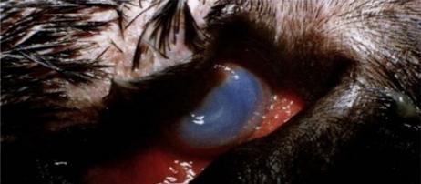
A San Bernard (detail introduction) occurred in the diamond -shaped eye, which manifested as a severe lower eyelid reversal, and the lower eyelids were turned inside the corner of the eye. The inner and outer corners of the upper eyelids are turned in the corner of the eye, the corneal matrix ulcer, the entire corneal edema and the neonatal and chronic keratitis accompanied by the edge blood vessels. Image source: Dog Eye Science color map (Kis. Barnett et al. Edited, the main translation of Wu Shouzheng)
The dog's normal eyes, right eyes
Two: cause
1 Developed outwardness is related to congenital inheritance, commonly in St. Bernard dogs, Hemati, Dandan dogs, Newfoundland dogs and bullfighting dogs. These dogs are often relaxed by the lower eyelids, which may be caused by obvious wide eyelids and lack of external contraction muscles. They are also the standard characteristics of such breed dogs.
2 The acquired veneer is caused by trauma or chronic inflammation. The formation of scars, fatigue, and bonding of eyelids and eyelid nerve injury can cause the disease.
3: Symptoms
The lower eyelid is reversed, the conjunctiva of the eyelid and the conjunctiva of the ball is red or dark red. Due to the long -term exposure of the conjunctiva, causing conjunctiva inflammation and tears, the inner eyes were wet.
Four: Treatment
For cases that are not serious outside the lower eyelids, chronic tear dogs do not need to be treated with surgery. Antibiotics and corticosterol eye drops can be used to reduce local stimulation and prevent infection. If the secretion is abnormally increased, chronic conjunctivitis and eyelid spasm occur, and it is suitable for surgical correction. There are many kinds of disease correction methods, but generally use WARTON-JONES eyelid formation, which is also V-Y technology. At 2-3mm under the edge of the lower eyelid, a deep-skinned V-shaped skin incision is made at 2-3mm. Then separate the subcutaneous tissue from the tip of the V -shaped incision, and gradually free the triangular skin. Then do a proper stealth separation under the wounding skin on both sides. From the V -shaped tip, make nodules sutured upwards, and move the leather flap on the side of the suture, until the lower eyelid margin of the valga is restored to the original state, and it is corrected. Finally, the remaining skin incision is sutured, and the original V -shaped line incision will become Y -shaped. Switching is commonly used for surgery, and the needle distance is 2mm, and the stitching thread is removed 10-14 days after surgery.
Five: Postoperative care
Antibiotic eye drops or eye ointment should be used after surgery, 3-4 times a day, and maintained for 5-7 days. Eliminate the conjunctivitis or corneal inflammation symptoms due to the secondary eyelids; at the same time, pay attention to animal scratching or friction, causing damage to the department.
