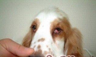
"Eyes are the windows of the soul." This is also very tried on the dog. Although humans cannot communicate with dogs language, dog's eyes and expressions can tell us what it is thinking. If the dog's eyes are sick, it is not only the dog must be very worried as the owner. This article mainly introduces a common dog eye disease-the cause of the third eyelid hyperplasia of dogs and symptoms and related diagnosis and treatment.
The third eyelid hyperplasia refers to the hyperplasia of the gland in the transient spine, and the outward turning over the transient membrane to free the edge, and a kind of eye disease from the inner corner of the eye, commonly known as "cherry eye".
The third eyelid is located in the inner corner of the eye, hiding behind the eyelids. Under normal circumstances, the naked eye can only be seen at most small edges. The main function of the third eyelid is to protect the cornea physiological, secrete tears and distribute tears on the surface of the eyeball, and can also clean the impurities of the cornea surface.

Cause
Congenital causes may attach congenital defects or dysplasia due to adrenal bases and orbital tissue or inter -gonad -to -cartilage connective tissue. It is generally believed that the transients or tubular mouths increased due to inflammatory products or small foreign bodies.
Symptoms
At the beginning of the disease, the conjunctiva of the patient's eyes was brightened and tears. Due to discomfort, some dogs scratched the affected eyes with fore limbs. One or two days later, the edge of the third eyelid of the eye was found to be the size of the mung bean, and the pink pink of the soybean bean is protruding from the inner corner of the eye. As the disease develops until the peanuts are large. Its surface is smooth, humid, and tough. In the early stage, because the prostate was smaller, he could often retract the eyelids by itself.
In the future, due to the increase of the prostate, it could not be shrunk, covering part of the cornea to affect the vision, and the color turned from the initial pink to purple -red.
Eye secretions change from the initial slurry to sticky purulentness. If it is bleeding due to scratching ulceration, secondary bacterial infections will cause local inflammation, secondary conjunctivitis and keratitis.
Diagnosis
Diagnosis can be made according to typical clinical symptoms.
Treatment
Surgery resection is the main treatment method. During the resection, pay attention to not damage the conjunctiva and instarial membrane. Washing the eye with antibiotic eye drops, such as Olinky Eye, or San Luwei's antibacterial eye drop, then clamped the proliferative glands with a wound pliers, and gently pulled the cartilage to the outside of the eye.
Use analgesic pliers between the hyperplasia and the cartilage and lock the clamping buckle. Use scissors to remove the proliferation above the hematopoietic tong, and then use the burning method to burn the hemostasis, release the hematopoietic pliers, and apply erythromycin in the corner of the eyes. ointment.
After surgery, antibiotic eye drops are dripped for 3-4 days, 5 times a day.
The third eyelid is removed. Without the cover of the eyelids, the eyeball will be exposed, the attack of foreign bodies, the eyeball will also be dry and uncomfortable, and embedding can reduce the incidence of later dry eye.
Summary
The third eyelid glands were not completely removed during the operation, but the part of the gland was properly retained, accounting for about 1/5 of the gland. According to clinical observations, no cases of secondary conjunctivitis keratitis were found, and better results were achieved.
There is also a clinical at a glance after being cured.
